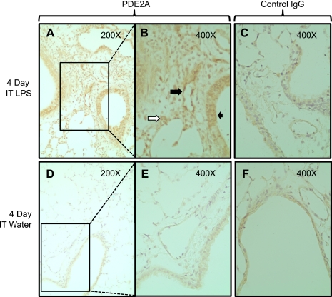
Fig. 2.
Lung immunohistochemistry for PDE2A expression 4 days following IT LPS (A and B) or water (D and E). Rectangles shown in A and D are shown in B and E under higher magnification. Large solid black arrow demonstrates endothelial PDE2A staining in a conduit pulmonary vessel. Small black arrow shows PDE2A staining in airway epithelium. White arrow demonstrates PDE2A staining in a macrophage. C and F: nonspecific IgG staining that was observed predominantly in airway epithelium in an LPS-independent fashion.

