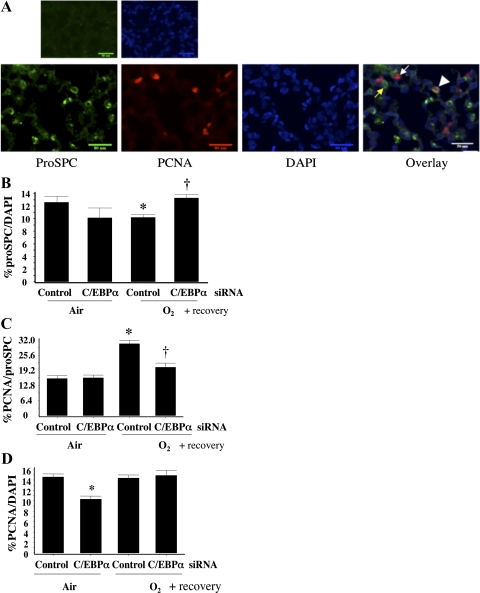
Fig. 7.
Evaluation of type II cell numbers and proliferating type II cells in control siRNA- and C/EBPα siRNA-injected lung after room air recovery. A: immunofluorescent staining of lung slides for pro-SP-C (indicator of type II cells), PCNA (indicator of overall proliferating cells), and 4′,6-diamidino-2-phenylindole (DAPI, indicator of total cells). Overlay shows cells positive for both pro-SP-C- and PCNA. Yellow arrow indicates pro-SP-C-positive cell, white arrow indicates PCNA-positive cell, and arrowhead indicates cell positive for both pro-SP-C and PCNA. B: pro-SP-C-positive cells as percentage of DAPI-positive cells. *P < 0.05 vs. control siRNA-injected lung (Control) in 72-h air exposure (Air). †P < 0.05 vs. Control in 72-h O2 exposure (O2). C: cells positive for both pro-SP-C and PCNA as percentage of DAPI-positive cells. *P < 0.05 vs. Control in Air. †P < 0.05 vs. Control in O2. D: PCNA-positive cells as percentage of DAPI-positive cells. Ten high-power fields were counted on each lung section at the distal alveolar epithelial level. *P < 0.05 vs. Control in Air. Values were obtained from an average of 3 lungs and an average of 10 high-power fields per lung.

