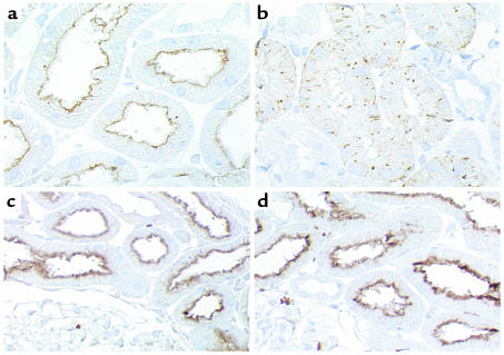
Figure 3.
Localization of cubilin (a and b) and megalin (c and d) in kidney cortex of normal (a and c) and affected (b and d) dogs. Sections were incubated with anti-dog cubilin (1:4,000). In normal dogs, cubilin is localized in luminal plasma membranes of kidney proximal tubules (a), whereas the labeling in affected dogs is intracellular and vesicular with no detectable receptor in the luminal plasma membrane (b), suggesting retention in an intracellular compartment. No difference in the labeling intensity or in the distribution of megalin is observed between normal (c) and affected (d) dogs when evaluated by immunocytochemistry using a sheep anti-rat megalin (1:10,000). ×600 (a and b); ×350 (c and d).

