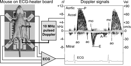
Fig. 2.
Illustration of the setup used to make noninvasive ECG and Doppler measurements showing an anesthetized mouse taped to electrodes on a temperature-controlled circuit board, Doppler and ECG signal processors, and a display of ECG and Doppler signals from the left ventricular inflow and outflow tracts. The 23 × 30-cm-printed circuit board contains 4 stainless-steel ECG electrodes, an array of 50 surface-mount resistors, a temperature sensor, a large ground plane, electrical connections to an ECG amplifier, and a temperature controller. Vel, velocity; Δf, change in frequency. Labeled on the Doppler tracing are the opening (o) and closing (c) of the mitral (m) and aortic (a) valves, peak ejection velocity (P) and acceleration (Accel), and peak-early (E) and late-filling (A) velocities. From these signals, one can obtain accurate timing of cardiac events such as preejection time, filling and ejection times, and isovolumic contraction and relaxation times as indexes of systolic and diastolic ventricular function [from Hartley et al. (25)].

