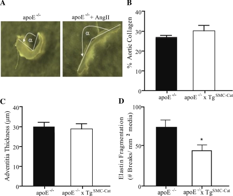
Fig. 4.
Opening angle of apoE−/− and apoE−/− + ANG II aortas and morphology of apoE−/− and apoE−/−xTgSMC-Cat aortas after ANG II treatment for 7 days. A: apoE−/− abdominal aortic opening angle (α) increased after ANG II treatment. B: collagen content decreased in the apoE−/−xTgSMC-Cat group and was unchanged in the apoE−/− after ANG II treatment. C: adventitial thickness in apoE−/−xTgSMC-Cat aortas decreased to the same level as apoE−/−. D: elastin fragmentation in the abdominal aortic wall of apoE−/− mice remained higher than in aortas from apoE−/−xTgSMC-Cat mice, with no change from baseline in either group. *P < 0.05; n = 3 for apoE−/−; n = 5–8 for apoE−/− + ANG II; n = 4 for apoE−/−xTgSMC-Cat; n = 5 for apoE−/−xTgSMC-Cat + ANG II.

