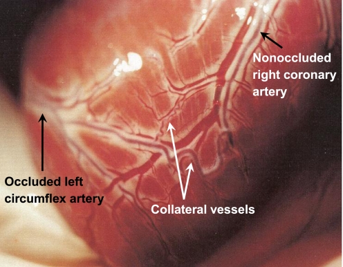
Fig. 1.
Representative photograph illustrating the apical view of a canine heart 4 mo following placement of an ameroid occluder around the proximal left circumflex coronary artery (entering from the left side of the photograph). Typical canine coronary collateral arteries are clearly visible on the epicardial surface, including both large (∼1 mm diameter) and smaller, tortuous arterial connections between a branch of the completely occluded left circumflex coronary artery and a branch of the nonoccluded right coronary artery. All animal protocols were in accordance with the “Principles for the Utilization and Care of Vertebrate Animals Used in Testing, Research and Training” and approved by the University of Missouri Animal Care and Use Committee.

