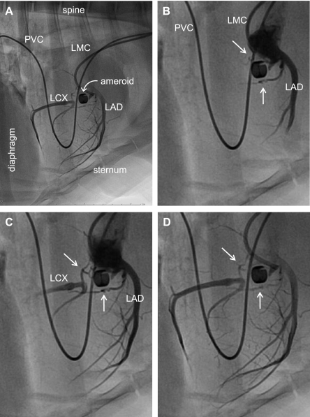
Fig. 2.
Angiography of porcine model of chronic coronary artery occlusion and collateral perfusion. Twenty-two weeks after surgical placement of an ameroid constrictor around the proximal left circumflex coronary artery, the animal was placed under general anesthesia. Hemodynamics were monitored throughout the procedure. Arterial access was obtained by surgical cutdown of the carotid artery. The left main coronary artery was catheterized with a 6F guiding catheter (Vista Brite Tip; Cordis) introduced over a 0.035-in. guidewire. Selective coronary angiography was performed with nonionic contrast (Oxilan 350; Guerbet). A: image of lateral view displaying the majority of the thoraci structure for perspective. B–D: regionally enhanced serial images emphasizing the cardiac silhouette to provide additional detail of coronary vasculature. White arrows in B–D identify collateral vessels supplying the left circumflex artery (LCX) distal to occlusion. LAD, left anterior descending artery; LMC, left main coronary artery catheter; PVC, pulmonary vein catheter for procedure not related to the data associated with this study. [Reproduced from Zhou et al. (89).]

