Fig. 1.
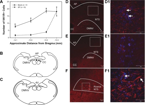
A: quantification of neurokinin-1 receptor (NK1R)-positive cell number in the nucleus tractus solitarii (NTS) in brain sections from four levels: the first three encompass the caudal NTS under the area postrema (AP), and the fourth section is in the rostral NTS. Only animals with at least a 70% decrease in NK1R-positive cells with substance P conjugated to saporin (SP-SAP) injected (dashed line) were considered lesioned. Values are means ± SE. *P < 0.05 from controls (solid line). B: location of caudal NTS as shown in D and E, approximately −13.8 mm from bregma. C: location of rostral NTS as shown in F and F1, approximately −13.3 mm from bregma. D: control animal injected with nonsense peptide conjugated to SAP (Blank-SAP). D1: higher magnification of caudal NTS of D. NK1R-positive cell bodies (arrows) and processes are present. E: animal injected with SP-SAP. E1: higher magnification of caudal NTS of E. No NK1R cell bodies were present; some processes were present. F: NTS rostral to SP-SAP injection site. F1: higher magnification of F. NK1R-positive cell bodies (arrows) and processes are present. For D and F, NTS is located inside white dashed line. Red staining denotes NK1R immunoreactivity, and blue the cell nucleus (4,6-diamidino-2-phenylindole). CC, central canal; DMNV, dorsal motor nucleus of the vagus; 4V, fourth ventricle.

