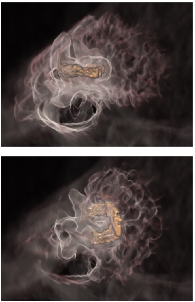
Figure 1.
Weighted distance field rendering for the lateral (top) and posterior (bottom) semicircular canals. The selected structures are rendered in high opacity orange, and nearby bony structures are rendered with a medium opacity white, fading opacity as the weighted distance from the structure increases.

