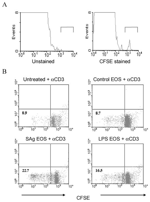
Figure 1.
Migration to the spleen and stimulation of T cell proliferation by intraperitoneally injected purified eosinophils. In panel A, 5 × 10⁶ carboxyfluorescein succinimide ester (CFSE)–labeled eosinophils were inoculated intraperitoneally into naive C57BL/6J mice. Spleens were recovered 36 h later, made into single-cell suspensions, and evaluated for the presence of CFSE-labeled eosinophils by use of a FACSCalibur flow cytometer. Representative data from 1 of 2 separate experiments are shown. In panel B, 5 × 10⁵ control eosinophils (indicated as “EOS” in the figure), eosinophils pulsed with Strongyloides stercoralis antigen (SAg), or eosinophils pulsed with lipopolysaccharide (LPS) were intraperitoneally inoculated into naive C57BL/6J mice. On day 10, spleen cells were labeled with CFSE and cultured in the presence of anti-CD3 antibody (αCD3) to demonstrate T cell proliferation. Representative data from 1 of 3 separate experiments are shown.

