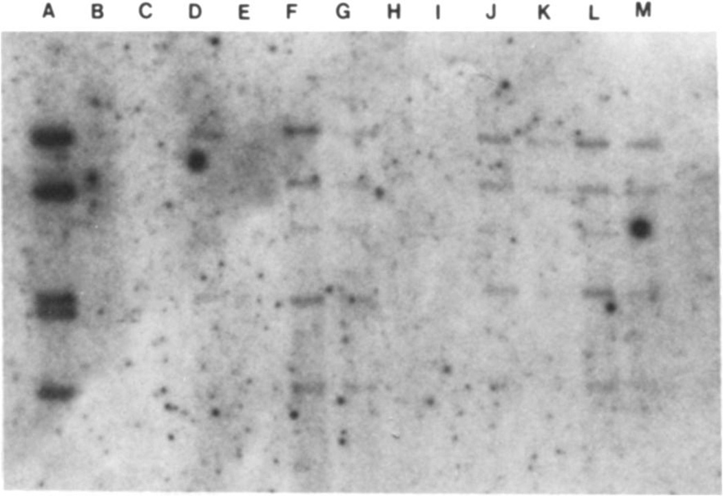
Figure 1.
Southern blot analysis of tumor tissue from organ transplant recipients with the EBV probe EcoRI B. Lane A contains the equivalent of 330 copies per cell of probe DNA in a sample of 106 cells. Lane D contains DNA from a lymphoid line containing 40 genome copies per cell; the DNA from 105 cells was loaded in this lane. Lane E contains DNA from an EBV-negative cell line. Lanes F and G contain 1 µg and 500 ng, respectively, of DNA from the lung biopsy of a heart transplant recipient (patient 13); lanes H and I contain 1 µg and 500 ng, respectively, of DNA from a lymph node of a heart-lung transplant recipient (patient 6); lanes J and K contain 1 µg and 500 ng of DNA from one tonsil, and lanes L and M contain 1 µg and 500 ng of DNA from the other tonsil of patient 5, a heart-lung transplant recipient. EBV DNA was found in tissues from patients 13 and 5 but not 6. Lanes G, K, and M each contain the DNA equivalents of 105 cells. The signals corresponding to these three samples represent the equivalent of ~40 copies per cell. Note that all samples contain a group of BamHI fragments expected to be present in EcoRI B probe.

