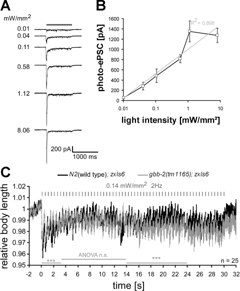
Fig. 6.
Graded responses of cholinergic MNs to increasing stimulus strength and different effects of low-intensity stimulus trains on ACh-evoked contractions in wild-type vs. gbb-2 mutant animals. A: photoevoked postsynaptic currents (photo-ePSCs) were measured in wild-type animals expressing ChR2 in cholinergic neurons (transgene zxIs6). Currents were recorded from voltage-clamped muscle cells (shown are representative single experiments) in response to a 1-s light pulse (470 nm, indicated by shaded bar) of the indicated light intensity. Peak inward currents were followed by a steady-state current that returned to baseline after the end of the stimulus. B: the peak currents were averaged (n = 6–7) and fitted with a single exponent. Values are means ± SE. C: wild-type or gbb-2(tm1165) animals with transgene zxIs6 were assayed as in Fig. 5B, with a light intensity of only 0.14 mW/mm2, and body contractions were quantified. Values are means ± SE; n = no. of animals analyzed. Two-factorial ANOVAs were used to analyze statistically significant differences (***P < 0.001) for time periods indicated by shaded bars.

