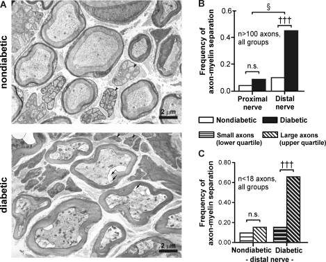
Fig. 4.
Axon-myelin separations are prevalent in distal axons from diabetic mice. A: electron micrographs of myelinated and unmyelinated axons in distal nerve segments from nondiabetic (top) and diabetic mice (bottom) stained with uranyl acetate and lead citrate (×3,430 magnification). Myelinated axons exhibited axon-myelin separations and numerous intracellular organelles compared with nondiabetic axons. Arrows highlight lipid-like material within axon-myelin separations. Arrowheads highlight some unmyelinated axons, which appeared more electron dense in diabetic nerves. B: frequency of axon-myelin separation in proximal versus distal nerve segments in nondiabetic and diabetic mice. C: frequency of axon-myelin separation in small-diameter axons (lower quartile) versus large-diameter axons (upper quartile). Quartiles were defined from the size distribution of all myelinated axons in nondiabetic or diabetic samples. †††P < 0.001 by a Kruskal-Wallis test followed by a post hoc Dunns multiple-comparison test between selected groups; §P < 0.05 by a Mantel-Haenszel test of homogeneity.

