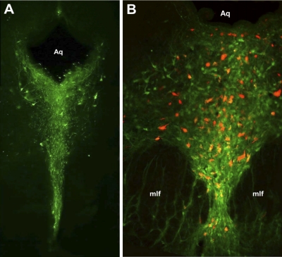
Fig. 2.
The majority of cells in the DRN that project to the NAc are serotonergic. A: coronal section of the PAG stained for tryptophan hydroxylase (TPH; green), demonstrating TPH-positive cells clustered in the DRN at approximately bregma −8.6. B: overlay image of retrogradely labeled DRN neurons that project to the NAc (red) and TPH-positive cells (green) at approximately bregma −7.6. In the population of backlabeled DRN-NAc projection neurons, 74.5% were colabeled for TPH (n = 1,378 backlabeled neurons from 6 rats).

