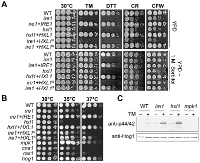
Figure 5. Functional analyses of C. neoformans HXL1 deletion and complementation.
(A) For sensitive test to UPR- and cell wall- stresses, strains were serially diluted and spotted on YPD media with or without exposure to ER stress agents and cell wall disturbing agents (0.075 µg/ml TM, 10 mM DTT, 5 mg/ml CR, 1.5 mg/ml CFW), without (top panel) or with 1 M sorbitol (bottom panel) and incubated at 30°C for 3.5 days. (B) For the thermosensitivity test, strains were spotted on YPD medium and incubated at 35°C or 37°C for 3.5 days. (C) Phosphorylation of Mpk1 induced by ER stress and loss of UPR function. Strains in early exponential phase were treated with TM (5 µg/ml) at 30°C for 2 hr. 30 µg of total protein was analyzed by Western blotting with phospho-p44/42-MAPK antibody (upper panel) for the phosphorylated Mpk1 (Mpk1-P) protein and anti-Hog1 antibody (lower panel) for the Hog1 protein as a loading control. Strains were: wild-type (H99), ire1 (YSB552), ire1+IRE1 complemented (YSB1000), hxl1 (YSB723), hxl1+HXL1 u complemented (YSB762), ire1+HXL1 u suppressed (YSB743), ire1+HXL1 s suppressed (YSB747), mpk1 (KK3), cna1 (KK1), ras1 (YSB53), and hog1 (YSB64).

