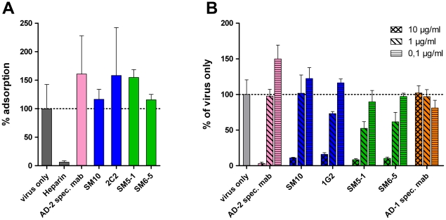
Figure 5. Mechanistic aspects of virus neutralization by Dom I and Dom II antibodies.
(A) Virus (m.o.i. 0.5) was incubated with the indicated antibodies (10 µg/ml for Dom I-, and Dom II-specific mabs, 5 µg/ml for the AD-2-specific mab C23, 2 µg/ml heparin) for 1 h at 37°C and cooled to 4°C. The virus/antibody mixture was added to HFF and incubated for 1 h at 4°C. Lysates were prepared and processed for quantitative real time PCR analysis. The virus only sample was set to 100% and used to calculate the remaining samples. (B) HFF were adsorbed with virus at a m.o.i. of 0.2 at 4°C for 1 h. Antibody at the indicated concentrations was added and the culture was shifted to 37°C. Extent of infection was analyzed 48 h later and calculated relative to the virus only control (100%). Color code of the antibodies according to their target structure on gB as shown in Fig. 3.

