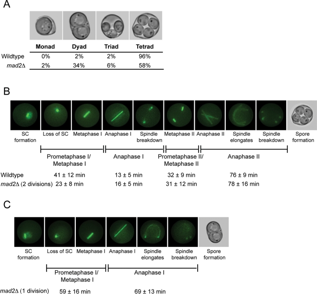
Figure 1:
Mad2 affects the duration of the meiotic cell cycle. (A) Wild-type and mad2Δ/mad2Δ sporulated cells were counted for the number of spores in each ascus. Nine hundred sporulated cells were counted in three biological replicates. (B, C) Synaptonemal complex (SC) formation and loss, and spindle assembly and disassembly, were visualized by expressing Zip1-GFP and TUB1-GFP and monitored using time-lapse fluorescence microscopy. The stages of meiosis were determined based on loss of Zip1 and spindle morphology, and the time of each stage was calculated (in minutes ± SD). (B) Still images from a representative movie of wild-type cells. One hundred cells of each genotype were counted. (C) Still images from a representative movie of mad2Δ cells that form dyads. Fifty mad2Δ cells that formed dyads were counted.

