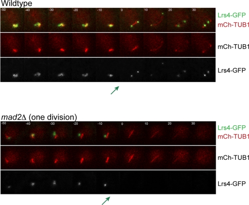
Figure 3:
In the mad2Δ cells that undergo one meiotic division, the monopolin complex does not bind sister kinetochores. (A) Time lapse images of meiosis in wild-type and 1 division mad2Δ/mad2Δ cells. Both strains are expressing Lrs4-GFP and Tub1-mCherry. The green arrow shows the point in which Lrs4-GFP leaves the nucleolus. One hundred of the wild-type cells, 100 of the 2 division mad2Δ cells, and 50 of the 1 division mad2Δ cells were analyzed.

