Figure 1:
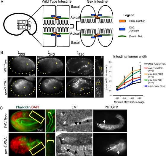
Arp2/3 is required during intestinal morphogenesis in C. elegans embryos. (A) Wild-type intestinal cells have apical junctions that recruit abundant F-actin; they pack together tightly and form a narrow intestinal lumen. Loss of Arp2/3 or WAVE/SCAR complex genes leads to the Gex, or gut on the exterior, morphogenesis phenotype. Gex intestinal cells recruit less actin at the apical junction; the cells are rounded and form an expanded intestinal lumen. (B) Live imaging to compare the intestinal lumen expansion in wild-type embryos and in embryos depleted of GEX pathway. Embryos are oriented with anterior to the left. The dlg-1::gfp transgene (Firestein and Rongo, 2001; Totong et al., 2007) is expressed at the apical junction of epithelial tissues during embryonic development, including the intestine (internal yellow brackets) and the epidermis (expression at the embryo surface). Yellow dots outline parts of the embryo not enclosed by epidermis. Yellow brackets indicate intestinal width. Right, a close-up of the intestine. Image contrast was enhanced equally to highlight intestinal width. Error bars show SEM. Asterisks indicate statistical significance, p < 0.05. (C) Left, phalloidin (green) was used to compare apical F-actin levels in wild-type and gex-3(RNAi) intestines. The yellow box around part of the intestine is amplified immediately to the right. Yellow brackets indicate intestinal lumen width. Middle, TEM of the intestine in wild-type and gex-3(RNAi) embryos. White arrows, apical adherens junctions; black arrows, microvilli; L, intestinal lumen. The white substance inside the lumen is the glycocalix. Right, the vha-6p::ph::gfp strain (labeled PH::GFP) was used to visualize cell membranes in wild-type and gex-3(RNAi) embryonic intestines. The split arrows illustrate the intestinal lumen width; the white single arrow points to lateral regions.

