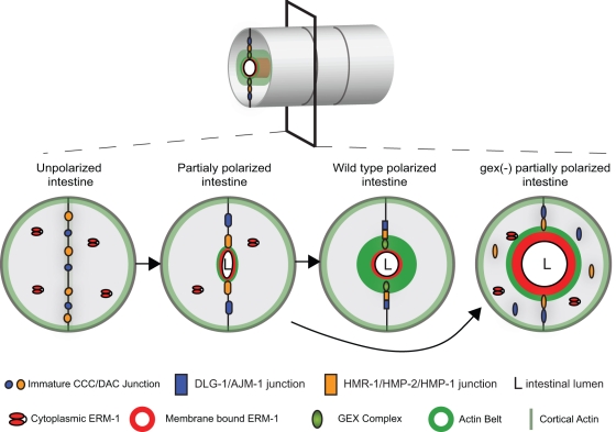
Figure 7:
A developmental model for the role of Arp2/3 and ERM-1 in apical junction formation during development. Cross–sections of the intestinal tube are shown in developing wild-type and Gex embryos. Polarization of intestinal cells requires the assembly of the apical domain, including apical localization of the junctions, ERM-1, the WAVE/SCAR complex, and F-actin. ERM-1 helps to establish the apical region by binding to cortical F-actin before the apical junctions have fully formed, by localizing WAVE/SCAR apically, and by repositioning the apical junction proteins apicolaterally. When Arp2/3 is present the correct levels of ERM-1 accumulate at the apical plasma membrane, and junctional proteins can accumulate at their correct apicolateral locations. In the absence of Arp2/3 or its activator, the WAVE/SCAR complex, ERM-1 levels are highly elevated. The elevated ERM-1 may displace junctional proteins from the apical domain, leading to decreased junctional proteins at the membrane, less apical F-actin, and an expanded lumen width. Taken together, these defects result in a cell that is only partially polarized.

