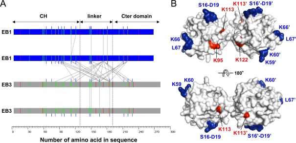
Figure 3:
Chemical cross-linking of EBs. (A) Intermolecular map of identified cross-links between lysine residues of EB1–EB1, EB1–EB3, and EB3–EB3 dimers. The proteins are depicted as bars, and cross-linked lysines (green vertical lines) are indicated by gray lines. Monolinks (blue vertical lines) and tryptic cleavage sites (arginine; red vertical lines) are marked. (B) Model of the CH domain pair. Red and blue residues were found to be cross-linked by DSS and to interact with microtubules (Slep and Vale, 2007), respectively.

