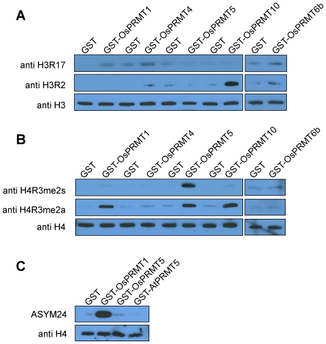
Figure 4. Site specificities of the OsPRMT1, OsPRMT5, OsPRMT6b, and OsPRMT10.
(A, B and C) Purified calf thymus core histones were either incubated with negative control, GST or GST-OsPRMT1, GST-OsPRMT5, GST-OsPRMT6b, GST-OsPRMT10 and GST-AtPRMT5 and were separated by 15% SDS-PAGE, followed by western blot analysis using anti-H3R2me2a, anti-H3R17me2a, anti-H4R3me2a, anti-H4R3me2s, and ASYM24 antibodies. An equal amount of the reaction mixtures were separated by the 15% SDS-PAGE, followed by western blot analysis using anti-H3 and anti-H4 antibodies, for equal loading (lowest panels in A, B and C). The antibodies are indicated to the left of each panel, whereas the corresponding PRMTs are represented above the upper panels.

