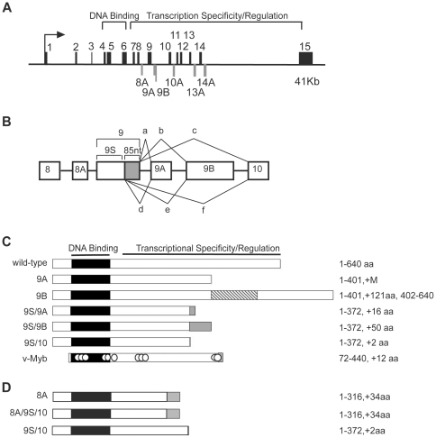
Figure 1. Alternative splicing in the c-myb gene.
(A) Structure of the human c-myb gene. The diagram shows the exon structure of the human c-myb gene, which spans more than 41,000 nucleotides. The arrow marks the major transcription initiation site and exons 1–15, which are included in the wild type version of c-Myb, are indicated above the line. The highly conserved DNA binding domain of c-Myb is encoded by exons 4–6, and the C-terminal domains that control transcriptional activity and specificity are encoded by exons 7–15, as indicated. The latter region also includes the 6 alternative exons, shown below the line. (B) Alternative splicing in exons 8–10. A cryptic splice donor site in exon 9 generates a short (9S) version lacking 85 nt (shaded). Both the long and short forms of exon 9 can be spliced to alternative exons 9A or 9B or to exon 10. (C) Diagrams of wild type c-Myb and the 9A, 9B, 9S/9A, 9S/9B and 9S/10 splice variants. The structure of the v-Myb protein encoded by Avian Myeloblastosis Virus is shown for comparison. The highly conserved Myb DNA binding domain is shaded black. The variant proteins lack the normal C-terminus of c-Myb and have unique regions, indicated by shaded boxes. The numbers on the right summarize the structures of the proteins with the included amino acids (aa) and the changes relative to wild type c-Myb. (D) Structures of the proteins encoded by splice variants 8A, 8A/9S/10 and 9S/10. These variants are described in the text. Labels and numbering are as in panel (C).

