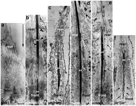
Figure 11. EM images of actin cones at late stage.
The indicated transgene was expressed in wild type (b) or myosin VI mutant (a, c–f) background. (a) GFP-M6FL (b) GFP-HNPMD (Gtail deleted) expressed in wild-type animal; (c) GFP-HNPMD expressed in a myosin VI mutant animal; (d) GFP-M6 mRRL in a myosin VI mutant animal, (e) GFP-M6mWKA in a myosin VI mutant animal; (f) no transgene (jar 1 mutant) Large arrow in (a) indicates the direction of cone movement, small arrows indicate areas near the cone center that lack actin filaments. mi, mitochondria; ax, axoneme. Bar, 1 µm.

