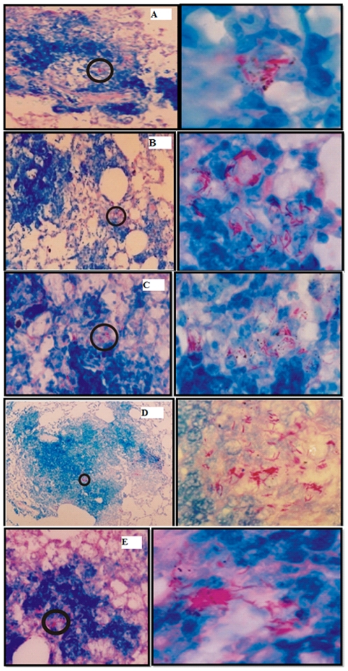
Figure 7. The histopathological study of lungs obtained from animals belonging to various immunized groups.
At 8 weeks post challenge, the lungs obtained from animals belonging to various immunized groups were fixed in formalin, to prepare them for section cutting. Subsequently, sections were stained with hematoxylin and eosin or Ziehl-Nelson stain to facilitate acid fast staining of bacilli as described in materials and methods section. Various experimental groups included in the study are: BCG (A), Sham control (B), Free Rv3619c (C), physical mixture of sham archaeosome and Rv3619c (D) and archaeosome entrapped Rv3619c (E). The panels on left side represent low power photomicrographs (200×; except D which is 100× of original picture) of sections while right side panel represents the high magnification (1000×) of the inset part of corresponding left panel. Acid fast bacilli can clearly be seen in higher resolution photomicrographs.

