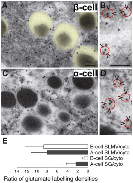
Figure 2. The glutamate concentration in SLMVs is much higher than in secretory granules, and lower in β-cell secretory granules than in α-cell secretory granules.
(A) Immunogold particles representing glutamate in β-cell cytoplasm are scarce over secretory granules (transparent yellow). (B) Close up showing glutamate gold particles in β-cell SLMVs (arrowheads, red circle). (C) Immunogold particles representing glutamate in α-cell cytoplasm. (D) Close up showing glutamate gold particles in α-cell SLMVs (arrowheads, red circle). Scale bars A–D, 100 nm. (E) Immunogold quantification shows that the secretory granule (SG)/cytosol (cyto) ratio (mean±SD) of net glutamate labelling (background subtracted, see Methods) is significantly lower in β-cells than in α-cells (p<0.05, n = 5 cells of each kind) and that the SLMV/cytosol ratio is much higher than the SG/cytosol ratio in both α- and β-cells (p<0.01, n = 5 cells of each kind) (Mann-Whitney-U test, two tails). The mean glutamate labelling density (average number of gold particles/µm2±SD) in α-cell granules was 33.6±14.9, whereas the value in α-cell cytosol was 21.1±4.6 (p<0.05, Mann-Whitney-U test, two tails). In β-cells the glutamate densities were 19.7±12.4 in the granules and 30.2±7.6 in the cytosol (p<0.05, Mann-Whitney-U test, two tails). From ultrathin test sections with conjugates containing known concentrations of glutamate, which were processed with the glutamate antibodies in parallel with the glutamate labelling of islet tissue, a relationship between the concentration of fixed glutamate and the gold particle density in islet tissue can be approximately estimated. In β-cells the approximate concentration of glutamate was estimated to be in the lower mM range (2–3 mM in secretory granules and cytosolic matrix, respectively).

