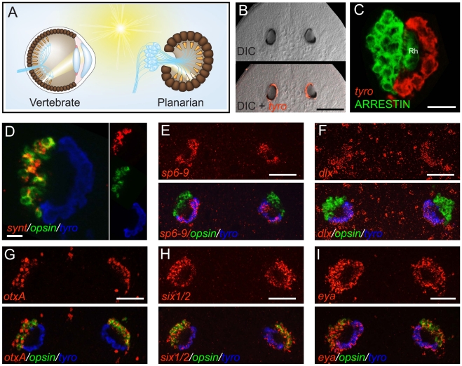
Figure 1. The planarian optic pigment cup expresses dlx and sp6-9.
Anterior is up in all images, and all eyes are shown at day 6 of regeneration except in (B). Fluorescent images are fluorescent in situ hybridizations (FISH) unless otherwise noted. (A) Schematic highlighting similarities between the vertebrate (left) and planarian (right) optic cup. Light-sensing organelles (orange), neuronal cell bodies and processes (blue), and pigmented cells of the optic cup (brown) are depicted. Neural circuitry of the vertebrate retina is highly simplified. (B) Smed-tyrosinase (tyro) is expressed in the optic (pigment) cup of an intact planarian. (C) Planarian photoreceptor neurons, labeled by an anti-ARRESTIN antibody [60] are adjacent to the pigment cup and extend rhabdomeres (Rh) into the cup lumen. (D) The optic cup does not express opsin or synaptotagmin (synt). (E–I) Expression of transcription factors in the planarian eye during head regeneration. sp6-9 (E) and dlx (F) are expressed in the optic cup. (G) otxA is expressed in the photoreceptor neurons, similar to the case for D. japonica [20]. six1/2 (H) and eya (I) are expressed in both the optic cup and photoreceptor neurons, similar to the case for other planarian species [3]–[4]. Scale bars, 100 µm (B, D), 50 µm (C, E–I).

