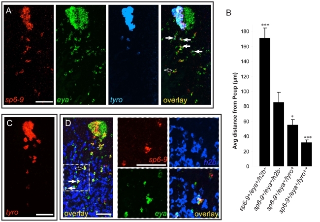
Figure 4. Optic cup trail cells exhibit a gradient of differentiation that correlates with distance from the cup primordium.
Anterior is up in all images, and all eyes and trails are in blastemas at day 6 of regeneration following decapitation. (A) sp6-9+/eya+ trail cells strongly expressing tyrosinase are close to the optic cup. (B) Quantification of distance of trail cells from the Pcup (mean ± s.e.m; n = 4 eyes for each category; significance by two-tailed t-test is shown relative to second bar (sp6-9+/eya+) ***P<.001, *P<.05). tyro++ indicates strong, non-granular signal. (C) Weak tyrosinase expression can be detected in cells far from the optic cup. (D) Some sp6-9+/eya+ cells express the proliferation marker histone h2b. Solid arrows show triple-positive cells and the open arrow shows double-positive cells. Scale bars, 50 µm.

