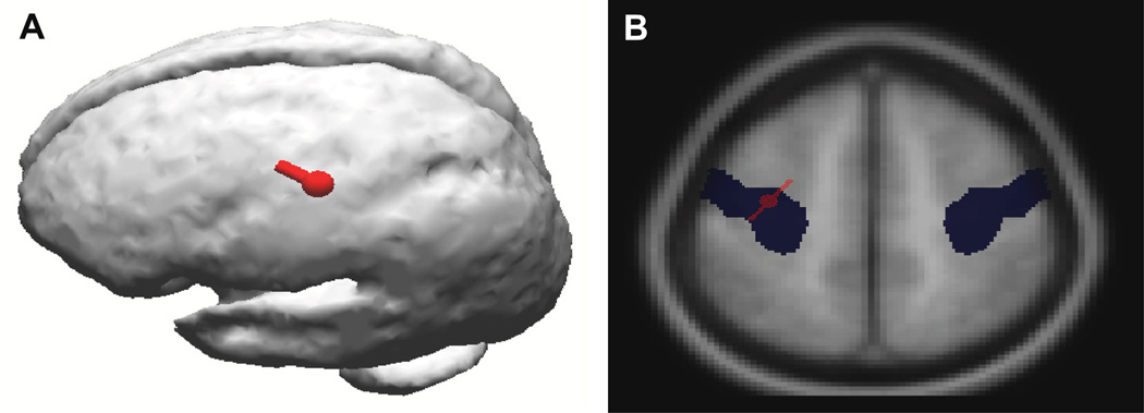
Figure 1.
A-(left) shows dipole localization (red) of the abnormal electrical activity (represented by a dipole) from the cortical myoclonus generation in a PD+Myoclonus subject from the right hand/wrist. This shows that the physiologic abnormality that produces the cortical myoclonus is a highly focal neocortical location. B-(right) shows that the same cortical myoclonus electrical activity (red) also overlaps the Talairach coordinates (blue) of the precentral gyrus (primary motor cortex) on MRI. These data provide evidence that the primary motor cortex is the location of the pathology that produces the cortical myoclonus in PD.

