
Figure 1.
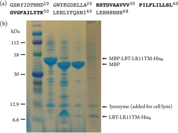
(a) Primary structure of the LBT-LR11TM-His6. The LR11 fragment is shown in bold, corresponding to residues 2132 to 2161 of the full-length protein. The LBT (lanthanide binding tag) is shown in italics. (b) SDS-PAGE results for the preparation of LR11 TM in native E. coli membranes. Lanes: 1, protein marker; 2, isolated E. coli membrane fraction; 3, thrombin cleavage of the sample in lane 2; 4, buffer washes of the sample in lane 3; 5, prepared membrane fraction for NMR experiments.