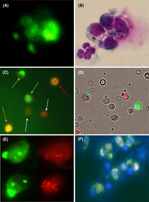
Fig. 1.
a Fluoromicrograph of a typical group of four epithelial antigen-positive cells (taken from Darb-Esfahani et al. 2009). Three of the cells display tiny caps, whereas the fourth exhibits strong staining, which was considered as intracellular staining. b Hematoxylin–eosin staining (HE) reveals that the intracellularly stained cell is a dying cell. In addition, a normal epithelial antigen-negative leukocyte is visible only in HE staining. c Fluoromicrograph of epithelial cells with green fluorescence, exclusively surface located including two vital epithelial antigen-positive cells with typical green caps (green arrows), adjacent to two faintly CD45-stained granulocytes (white arrows) and a bright CD45 stained vital lymphocyte (red arrow) in addition to a dead PI-positive cell (orange arrow). d Overlay of a fluoromicrograph with transmitted light. Note, the red 7AAD-positive fluorescing nucleus of a dead normal leukocyte in the neighborhood of the green fluorescing CETC and a red and green fluorescing dying CETC. e Two CETC from the same patient using the green and red filter, respectively. In both cells, the typical green cap of the EpCam, and in addition, the two green signals of the CEP17 alpha DNA probe can be identified. The upper cell carries only two HER2/neu signals, whereas in the lower cell more than 10 signals can be detected. f Fluoromicrograph overlay of the HER2/neu FISH analysis of a group of tumor cells from a breast cancer patient: besides the normal blood cells (nuclear stain was DAPI, only the nuclei are visible), one can see a group of tumor cells staining with typical green caps with the FITC-labeled anti-EpCAM. These cells carry a varying quantity of HER2/neu amplifications (pink-red dots in the cell nucleus)

