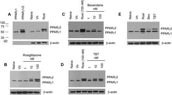
FIG. 5.
TBT induces expression of PPARγ in a bone marrow MSC model. BMS2 cells were allowed to become confluent and then treated with Vh (DMSO, 0.1%), rosiglitazone, bexarotene, or TBT (1–100 nM) (A–D) or rosiglitazone, bexarotene, and TBT (50nM) (E) in the presence of insulin (0.5 μg/ml) for 4 days. Whole cell lysates were prepared and analyzed for PPARγ expression by immunoblotting. Identity of the bands was confirmed by comparison with in vitro translated mouse PPARγ1 and PPARγ2 (A). β-actin expression was determined to assure equal protein loading. Data are representative of three independent experiments.

