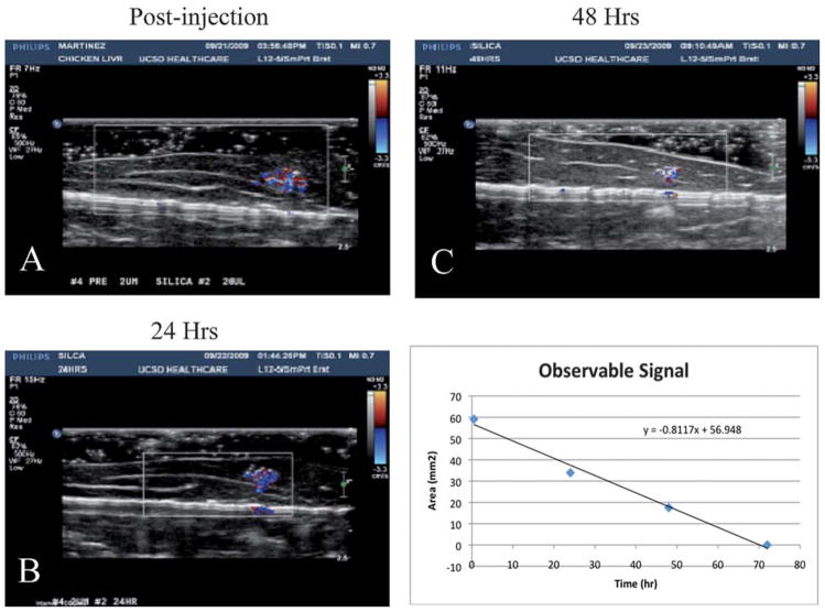
Fig. 5.
CDI sonograms of 100 μg of PFC-filled microshells injected in a tissue phantom imaged at 24 h (A) 24 h, (B), 48 h, and (C) 72 h. The signal decreases over time, but is still easily visible after 48 h. (D) CDI observable signal vs. imaging delay time. A fit of the data shows that the microshells have a half-life ~24 h.

