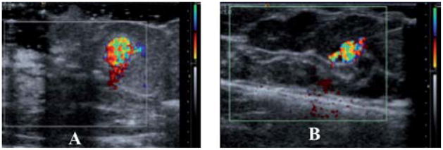
Fig. 6.
CDI images of 100 μL of PFC filled nano and microshells in human masectomy tissue: (a) after injection of 4 × 10¹⁰ nanoshells. (b) After injection 8 × 10⁸ microshells The nanoshells image (A) is magnified ~2.5× and thus in this comparison appears to be larger. An accurate measure the DCI area can be found in Table 1.

