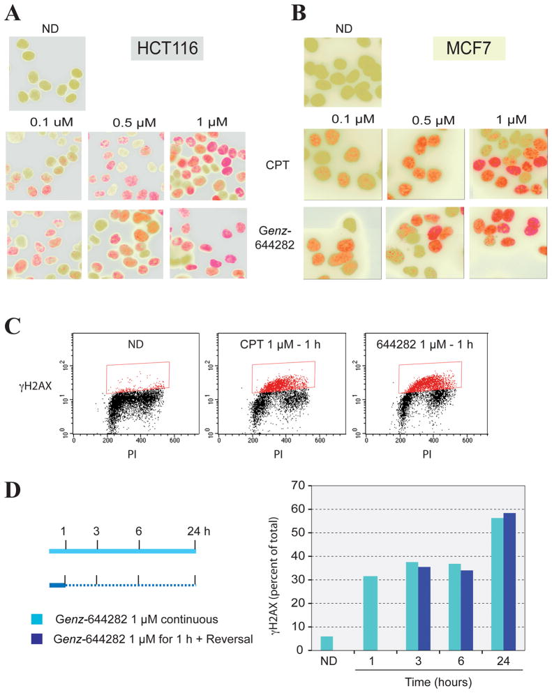
Figure 4. Generation and persistence of γH2AX foci in MCF7 and HCT116 cells in response to Genz-644282 treatments.
A and B) Immunofluorescence microscopy of γ-H2AX foci formation in HCT116 and MCF7 cells. Cells were treated with Genz-644282 or CPT for 1 hour. Accumulation of γ-H2AX is shown in red, nuclei are shown in green. C) Comparison of γ-H2AX-positive cells induced by CPT or Genz-644282 as analyzed by flow cytometry (PI: propidium iodide). D) Percentage of γ-H2AX-positive cells induced by continuous treatment with 1 μM Genz-644282 or 1 hour of treatment followed by incubation in compound-free medium for the indicated times. HCT116 cells were treated as shown in the left panel. Cells were fixed and incubated with γ-H2AX antibody and PI. The right panel depicts a quantitative comparison of γ-H2AX-positive cells induced by continuous compound exposure versus a 1-hour treatment followed by incubation in compound-free medium.

