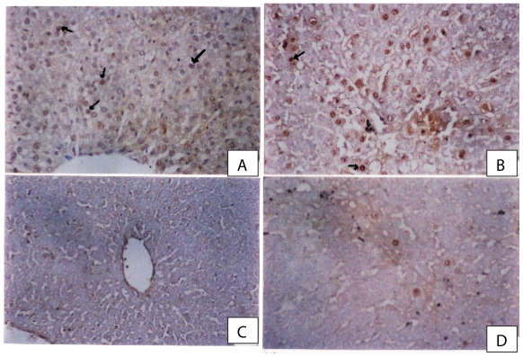
Figure 4.
(A) A photomicrograph from a section of control rat liver showing normal hepatocytes with few PCNA positive nuclei (arrows) (PCNA immunostaining × 300). (B) A photomicrograph from a section of rat liver given CCl4 showing prominent increase in the number of PCNA positive nuclei (proliferating cells) in regenerated cells in necrotic areas around central veins (PCNA immunoperoxidae × 300). (C) A photomicrograph from a section of rat liver given CCl4 and a combination of ribavirin (60 mg/kg)and silymarin showing marked reduction in the number of PCNA positive nuclei especially in peripheral zones compared with sections from rats treated with CCl4-olive oil (PCNA immunostaining × 300). (D) A photomicrograph from a section of rat liver given CCl4 and ribavirin at 90 mg/kg showing few number of PCNA positive nuclei in hepatocytes adjacent to the central vein (PCNA immunostaining × 300).

