Figure 1.
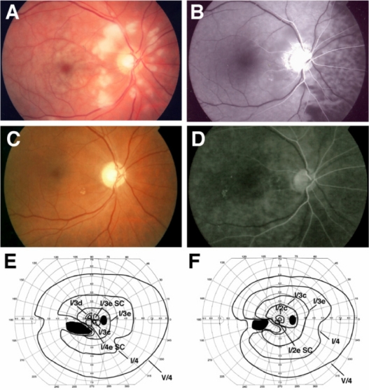
Clinical findings for Case 1. Fundus photographs and fluorescent angiograms before treatment. There are many cotton wool patches surrounding the optic disc and retinal whitening surrounding the fovea (A). Fluorescein angiography shows nonperfused areas of retinal arterioles in the areas corresponding to the areas of cotton wool patches. A delay in the choroidal filling in the nasal area and arm-to-retina time (32 seconds) was found (B). Fundus photograph (C) and fluorescent angiogram (D) after treatment. Optic disc is slightly pale, and defects of the nerve fiber layer can be seen (C). Fluorescein angiography shows improvement of the retinal circulation (D). Arm-to-retina time was 15 seconds. Goldmann kinetic perimetry before (E) and after (F) treatment. There is a large absolute scotoma in the lower nasal field and paracentral scotoma before treatment (E). The paracentral scotoma is decreased after treatment (F).
Note: Black indicates absolute scotoma and gray relative scotoma in E and F.
Abbreviation: SC, scotoma.

