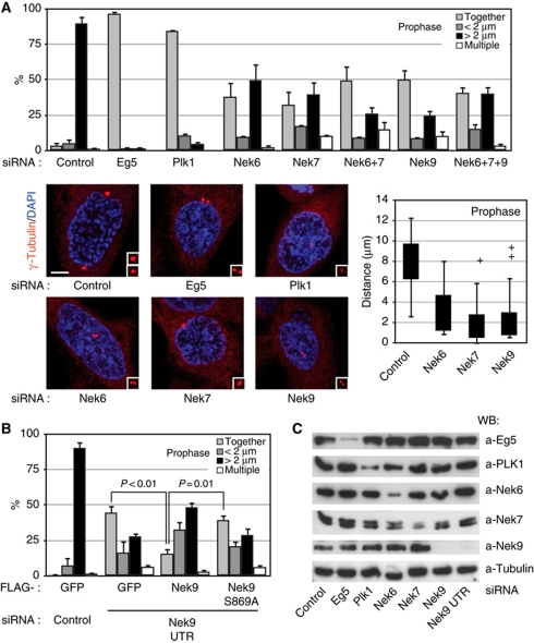
Figure 4.
Plk1, Nek9, Nek6, Nek7 and Eg5 are necessary for normal centrosome separation in prophase. (A) HeLa cells were transfected with the indicated siRNAs, and after 24 (Eg5, Plk1) or 48 (control, Nek6, Nek7, Nek9) hours, fixed and stained with antibodies against γ-tubulin (red) and DAPI (blue). Cells showing condensed chromosomes and intact nuclei (assessed by the shape of the DNA and a γ-tubulin exclusion from the nucleus) were scored as in prophase (these cells were 100% positive for histone H3[Ser10] phosphorylation, thus confirming the cell-cycle phase assignation, data not shown). The percentage of prophase cells showing two unseparated centrosomes (together), two centrosomes separated <2 μm (<2 μm), fully separated centrosomes (>2 μm) or more than two centrosomes (multiple centrosomes) is shown in the upper graphic (mean±s.e.m. of three independent experiments; ∼50 cells counted in each experiment). Additionally, the distribution of distances from the centre of the duplicated centrosomes in each case is shown as a box plot (boxes show the first and third quartiles, whiskers mark minimum and maximum values unless these exceed 1.5 × interquartile range and crosses correspond to outliers; 20 cells counted for each experimental condition). Representative examples of the observed phenotypes are shown (bar, 5 μm). In each case, insets show magnified centrosomes. (B) HeLa cells were cotransfected with either control or Nek9 3′ UTR siRNAs plus expression plasmids for the indicated FLAG-tagged proteins, and 48 h latter processed and FLAG-positive cells scored as in (A) (mean±s.e.m. of three independent experiments; ∼40 cells counted in each experiment; statistical significance was determined using the standard Student's t-test). Levels of endogenous and recombinant Nek9 as determined by western blot are shown in Supplementary Figure S4C. (C) Efficiency of the different RNAi treatments used in (A) or (B) as determined by western blot of total cell extracts.

