Figure 6.
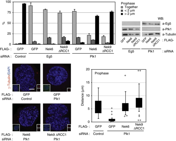
Active Nek9 and Nek6 can rescue Plk1 but not Eg5 downregulation in prophase centrosome separation. HeLa cells were transfected with control, Eg5 or Plk1 siRNAs plus the indicated plasmids and processed as in Figure 5 (Nek9ΔRCC1, Nek9[Δ346–732]). The percentage of FLAG-positive prophase cells showing two unseparated centrosomes (together), two centrosomes separated <2 μm (<2 μm) or fully separated centrosomes (>2 μm) is shown in the upper graphic (mean±s.e.m. of three independent experiments; ∼40 cells counted in each experiment). The effect of the different transfections on the levels of Plk1 and Eg5 is shown (right). Lower panels show representative examples of the observed phenotypes (anti-γ-tubulin plus DAPI staining, bar, 5 μm; insets show the same field stained with anti-FLAG plus DAPI) and a box plot of the distribution of distances from the centre of the duplicated centrosomes in FLAG-positive cells (as in Figure 4; 30 cells counted for each experimental condition).

