Figure 8.
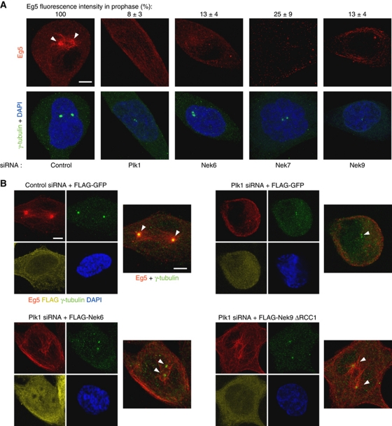
Plk1, Nek9, Nek6, Nek7 are necessary for centrosome recruitment of Eg5 during prophase. Active Nek9 and Nek6 can rescue Plk1 downregulation in prophase Eg5 recruitment. (A) HeLa cells were transfected with the indicated siRNAs, and after 24 (Plk1) or 48 (control, Nek6, Nek7, Nek9) hours, fixed and stained with antibodies against Eg5, γ-tubulin and DAPI. Representative examples of Eg5 (red) distribution in prophase cells are shown. Insets show the same field stained with γ-tubulin (green) and DAPI (blue). Centrosomal accumulation of Eg5 is noted with arrowheads. Bar, 5 μm. The efficiency of the different RNAi treatments can be seen in Figure 4C. Centrosomal Eg5 fluorescence intensity was quantified with ImageJ software on images acquired under constant exposure, using a circular area of 2 μm diameter surrounding a single centrosome (identified by γ-tubulin staining; an adjacent area of the same dimensions within each cell was quantified and subtracted as background). Results are expressed as a percentage of the intensities measured in control cells±s.e.m. (three independent experiments; >20 centrosomes counted in each experiment). (B) HeLa cells were cotransfected with either control or Plk1 siRNAs and expression plasmids for the indicated proteins and after 24 h fixed and stained with antibodies against Eg5, γ-tubulin and DAPI (Nek9ΔRCC1, Nek9[Δ346–732]). After incubation with labelled secondary antibodies, FLAG was detected with Fab-prelabelled anti-FLAG (see Materials and methods). Representative examples of Eg5 distribution in prophase cells are shown. Images show the same field stained with Eg5 (red), γ-tubulin (green), FLAG (yellow) and DAPI (blue), and a composite of Eg5 (red) plus γ-tubulin (green). Centrosomal accumulation of Eg5 is noted with arrowheads. Bar, 5 μm.

