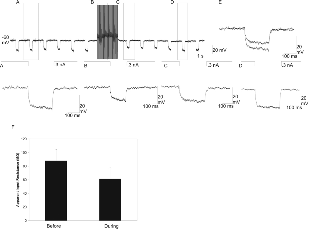
Fig. 5.
Decrease in the apparent input resistance in an nRt/PGN neuron with HFS delivered to the A1 lamina ~600 µm posterior to the recording electrode. Intracellular recording from a nRt/PGN neuron during HFS while hyperpolarizing square-wave current pulses (100–300 ms, 0.3 nA, 1 Hz) are delivered intracellularly to the recorded neuron. (A) Before HFS, the 0.3 nA hyperpolarizing DC resulted in membrane hyperpolarization. (B) Magnification of the labeled portion of the top trace with the stimulation artifacts manually removed. The hyperpolarizing current pulse resulted in significantly less membrane hyperpolarization compared to the pre-HFS hyperpolarizing current pulse. (C) Enlargement of the top trace immediately following HFS. The hyperpolarizing current pulse resulted in significantly less membrane hyperpolarization compared to the pre-HFS hyperpolarizing current pulse. (D) Magnification of top trace following HFS, showing return to baseline apparent input resistance after ~4 seconds post HFS. (E) Overlay of the membrane response to the hyperpolarizing current pulses before and during HFS. (F) graph of the mean ±SEM of the apparent input resistance before and during HFS (87.9 ± 14.8 MΩ and 61.3 ± 15.4 MΩ, respectively; n = 5 nRt/PGN neurons, P < 0.001).

