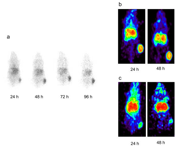
Figure 4.

Validation of panitumumab F(ab')2 as an imaging agent of HER1 positive tumors. (a) γ-Scintigraphy was performed with mice bearing LS-174T s.c. tumor xenografts. Following i.v. injection with approximately 100 μCi of 111In-CHX-A"-panitumumab F(ab')2, mice were imaged over a 4-d period. Positron-emission tomograhic (PET) using 86Y-CHX-A"-panitumumab F(ab')2. (b) Mice bearing s.c. LS-174T xenografts were injected i.v. with (50-60 μCi) of 86Y-CHX-A"-panitumumab F(ab')2 and imaged 1 and 2 days post-injection of the RIC. (c) Specificity was demonstrated by co-injecting 100 μg of panitumumab with the RIC and blocking uptake of the RIC by the tumor.
