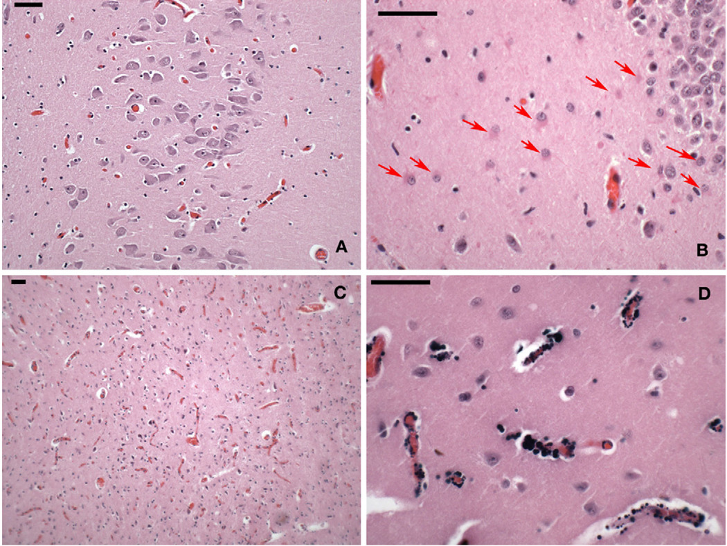
Figure 2.
Photomicrographs of postmortem brain tissue of the 19-year-old decedent (hematoxylin and eosin stain). Each scale bar = 50 µm. (A) CA1 sector of hippocampus, the area of the brain most vulnerable to systemic hypoxia-ischemia, with no indication of loss of neurons or gliosis. (B) CA4-subplate region of the hippocampus with a band of reactive astrocytes below the dentate layer of the hippocampus (arrows). (C) Hyperemia of the superior colliculus, also seen in the globus pallidus. (D) Granular perivascular calcification in putamen, uncommon in this age group and location.

