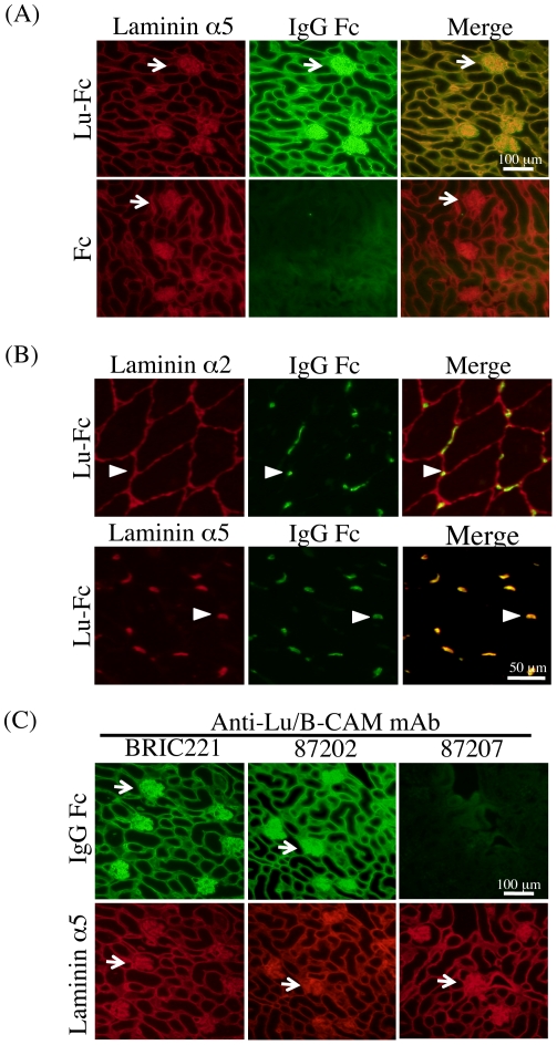
Figure 1. Lu/B-CAM binding assay on tissue sections.
(A) Binding of Lu-Fc to laminin α5 in adult mouse kidney sections. Sections were incubated with Lu-Fc (upper panel) or Fc (lower panel) at room temperature for 1 hour. Endogenous laminin α5 and bound Lu-Fc were detected with anti-laminin α5 domain LEb/L4b and anti-human IgG, as indicated. Merged pictures showed that laminin α5 and Lu-Fc co-localized at basement membranes. Arrows indicate glomeruli, laminin α5-rich structures. Bar, 100 µm. (B) Binding of Lu-Fc to laminin α5 in adult mouse skeletal muscle sections. Endogenous laminin α2 (upper panel) and α5 (lower panel) were detected with antisera. Bound Lu-Fc was detected with an antibody against human IgG as described above. Lu-Fc only bound to the basement membranes containing α5, primarily neuromuscular junctions. Arrowheads indicate neuromuscular junctions. Bar, 50 µm. (C) Inhibitory effects of anti-Lu/B-CAM antibodies on Lu-Fc binding to laminin α5. After preincubation with anti-Lu/B-CAM antibodies, Lu-Fc was used in the binding assay on tissue sections. Antibody 87207 inhibited the binding of Lu-Fc to endogenous laminin α5.

