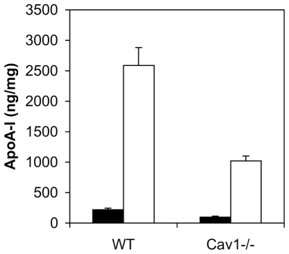
Figure 3. ApoA-I binding to NDR and non-raft membranes in WT and Cav1−/− MEFs.
ApoA-I binding to light (fractions 2–5, filled bars) and heavy (fractions 8–12, hollow bars) membranes was quantified by scintillation counting. ApoA-I (50 µg/mL) was bound at 4°C on ice and MEFs were fractionated into NDR and non-raft membranes as described for Fig. 1E. Data are average ± standard deviation of five experiments.

