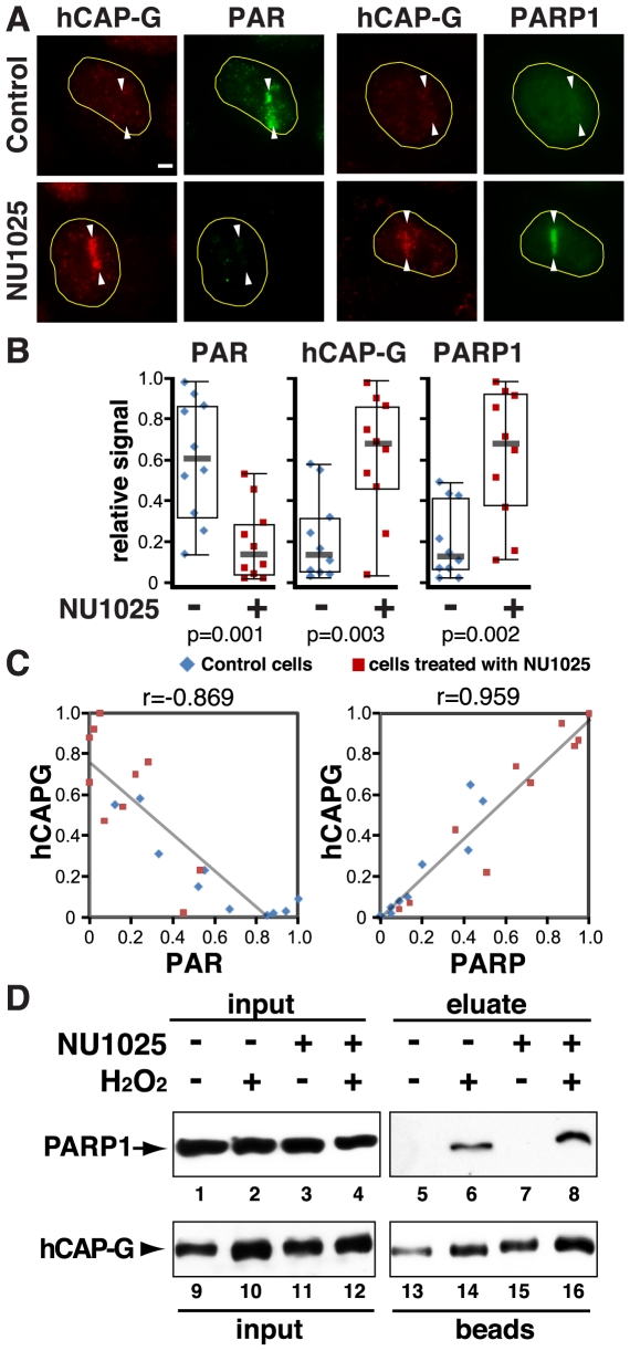
Figure 2. PARP1 inhibition enhances and prolongs PARP1 and condensin I accrual at the damage sites, and does not impair the condensin I-PARP1 interaction.
(A) Cells were damaged in the presence or absence of NU1025 (100 µM) and fixed at one hour after irradiation. Cells were stained with antibodies specific for PAR, hCAP-G, and PARP1 as indicated. (B) Comparison of the relative signals of each antibody staining in the control DMSO-treated and NU1025-treated cells as in (A). Ten cells were measured in each group. The relative signal was calculated based on the highest value of fluorescent signal in each antibody group and displayed as boxplots with medians. The p values were generated using a t-test and are shown at the bottom. (C) Correlation of the hCAP-G/PAR and hCAP-G/PARP1 co-staining in individual cells is plotted with trend lines. Pearson's r values are shown at the top. (D) Co-IP of PARP1 with condensin I from HeLa nuclear extracts using anti-hCAP-G antibody with or without NU1025 in the presence or absence of damage.

