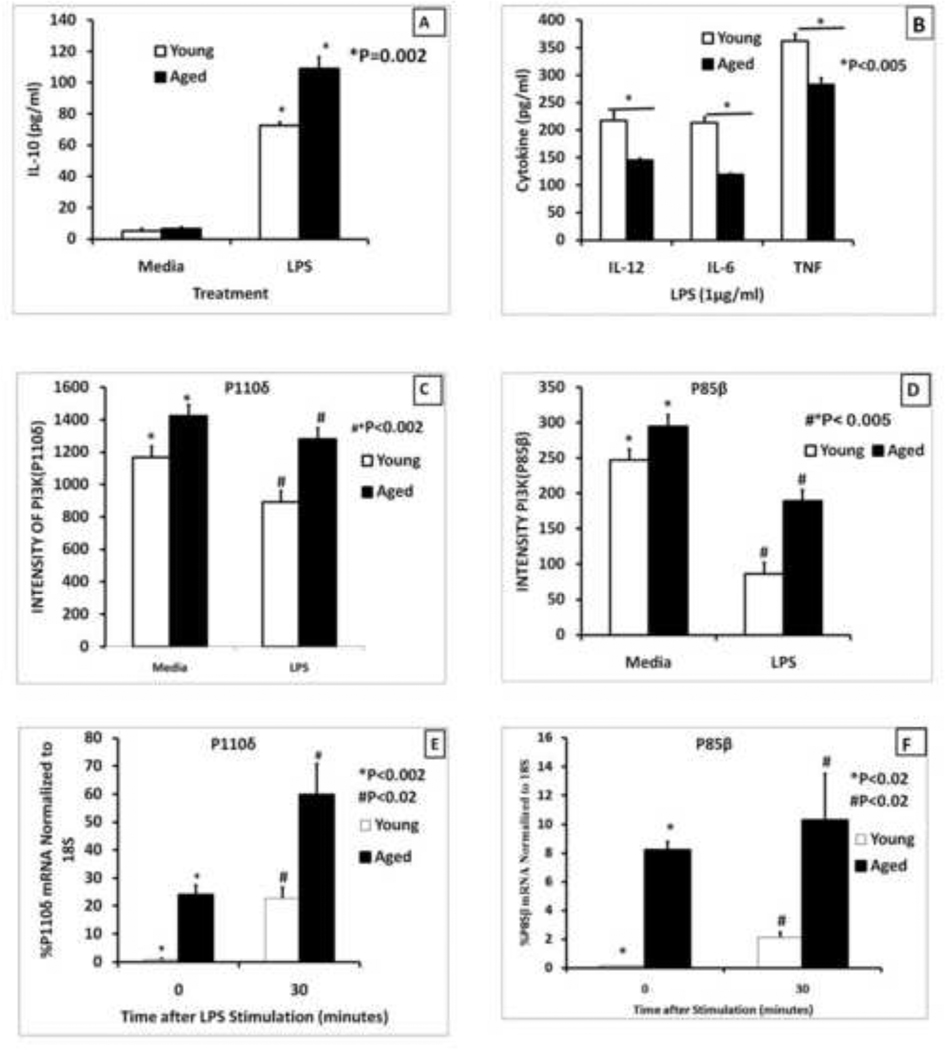
Figure 1. Age-associated changes in cytokine production and levels of PI3K subunits in splenic macrophages stimulated via TLR4.
Splenic macrophages (2.5×10⁵ cells/ml) from young (2–4 months) and aged (20–24 months old) mice purified by negative selection using an AUTOMACS cell separator were stimulated with LPS (1µg/ml) for 24 hours (Panels A and B) and the supernatants were collected and analyzed for IL-10 (Panel A) or pro-inflammatory cytokines, IL-12(p40), IL-6, and TNF-α (Panel B ). The results are shown as mean +/− SE of duplicate determinations from triplicate cultures, and are representative of three independent experiments. The symbol * indicates the statistical significance of differences in cytokine secretion between the young and the aged.
Panels C and D represent levels of RNA for p110δ and p85β subunits derived from microarray analysis performed on gene expression in young and aged macrophages as reported in Chelvarajan et al.(R. L. Chelvarajan, et al., 2006). QRT-PCR analysis of mRNA is shown in panels E and F for P110δ and p85β subunits respectively of PI3 kinase in purified splenic macrophages from young and the aged. The Ct values for each probe were normalized to the 18S RNA. The symbols * and # indicate the statistical significance of differences in RNA expression between the young and the aged.

