
Figure 8. Age-associated cytokine defect in TLR-2 and HKSP activated macrophages is due to a defect in the AKT-GSK-3 signaling axis.
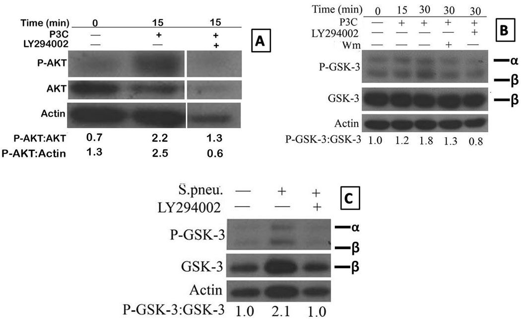
Purified aged splenic macrophages were pre-treated with either LY294002 or wortmannin for 1 hour and then stimulated with Pam-3-CSK4 (Panels A and B) or HKSP (2×10⁸ CFU/ml) (Panel C) for 15 or 30 minutes, respectively. The blots were probed for p-Akt (Panel A), p-GSK-3αβ (Panels B and C) and later probed for total Akt (Panel A), total GSK-3β (panels B and C) and actin after stripping. The numbers represent densities of bands normalized to total AKT, actin or GSK-3 with the values for unstimulated aged macrophages set to 1. Results are representative of two independent experiments.
The final figure in A is a composite of blots for P-Akt, Akt and actin for aged samples run on the same membrane that was stripped and reprobed. The intervening space (between second and third lane) was for lanes loaded with lysates from other time points or treated with other inhibitors but were deleted to focus on the effect of P3C on Akt activation at 15 minutes, which was maximum in this experiment.