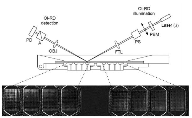
Figure 1. Oblique-incidence reflectivity difference (OI-RD) scanning microscope demonstrates that H. pylori binds to the Leb antigen also in a BabA-independent manner.
OI-RD scanning microscope equipped with a combination of a y-scan mirror and an x-scan linear stage. A 1 in × 3 in glass slide printed with 8 microarrays, each over an area of 3 mm × 4.5 mm, was assembled with a fluidic system with 8 chambers, each of which contained a microarray (A). The fluidic system was mounted on the x-scan linear stage. PEM: photo-elastic modulator. PS: phase shifter. FTL: encoded scan mirror for y-scan. OBJ: objective lens. A: polarization analyzer. PD: photodiode detector. The OI-RD image displays eight protein microarrays, each comprised of 315 bovine serum albumin (BSA) spots (see Supplementary Information, Text S1).

