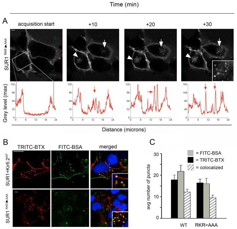
Figure 3. SUR1RKR→AAA expressed alone undergoes endocytosis.
(A) Live cell imaging of COSm6 cells expressing BTX tag-SUR1RKR→AAA. BTX tag-SUR1RKR→AAA expresses at the plasma membrane independently of Kir6.2 (acquisition start capture, scale bar 5 μm). Time-lapse imaging shows an increased number of fluorescent puncta inside the cells over 30min representing endocytosed BTX tag-SUR1RKR→AAA (10-30min movie screen captures, white arrowheads and inset). Internalization was analyzed using line scan and is illustrated in the graphs below (for details refer to Materials and Methods). (B) COSm6 cells expressing WT channels (BTX tag-SUR1+Kir6.2; upper panel) or BTX tag-SUR1RKR→AAA (lower panel) were labeled with TRITC-BTX and chased with FITC-BSA present in the medium for 30 min. Co-localization of WT channels and SUR1RKR→AAA (red, scale bar 10 μm) with FITC-BSA (green) confirmed visualized channels were intracellular (merged pictures, far right, insets, scale bar 10 μm). (C) Quantitative analysis showed that the average number of internalized BTX-labeled puncta was similar between BTX tag-SUR1RKR→AAA and WT channels as well as the number of BTX-stained vesicles colocalized with the endocytic marker FITC-BSA (BTX-labeled, black bar: SUR1RKR→AAA =16.5±1.7 and WT =16±2.4; colocalized FITC-BSA, striped bar: SUR1RKR→AAA =9.5±1.2 and WT=12.2±1.3, N=2, n>20 per condition) indicating similar internalization behavior for BTX tag-SUR1RKR→AAA and WT channels.

